45 eye diagram with labels and functions
Human Ear: Structure and Functions (With Diagram) - Biology Discussion The sound waves are collected by the external ear up to some extent. They pass through the external auditory meatus to the tympanic membrane which is caused to vibrate. The vibrations are transmitted across the middle ear by the malleus, incus and to the stapes bones. The latter fits into the fenestra ovalis. The Eye Diagram: What is it and why is it used? Here, the bit sequences 011, 001, 100, and 110 are superimposed over one another to obtain the example eye diagram. The eye diagram takes its name from the fact that it has the appearance of a human eye. It is created simply by superimposing successive waveforms to form a composite image. The eye diagram is used primarily to look at digital signals for the purpose of recognizing the effects of distortion and finding its source.
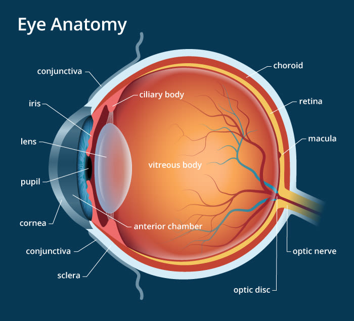
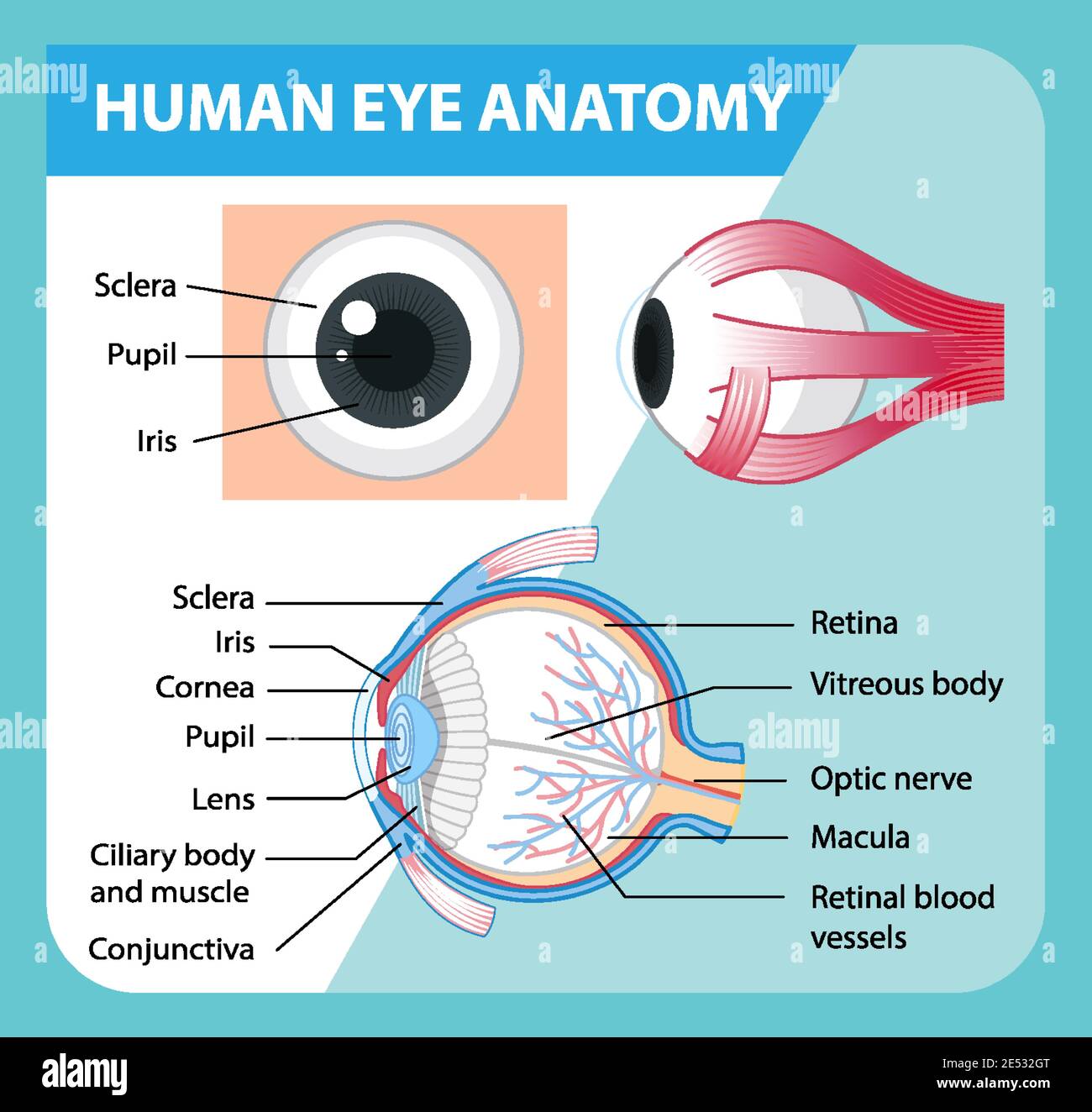
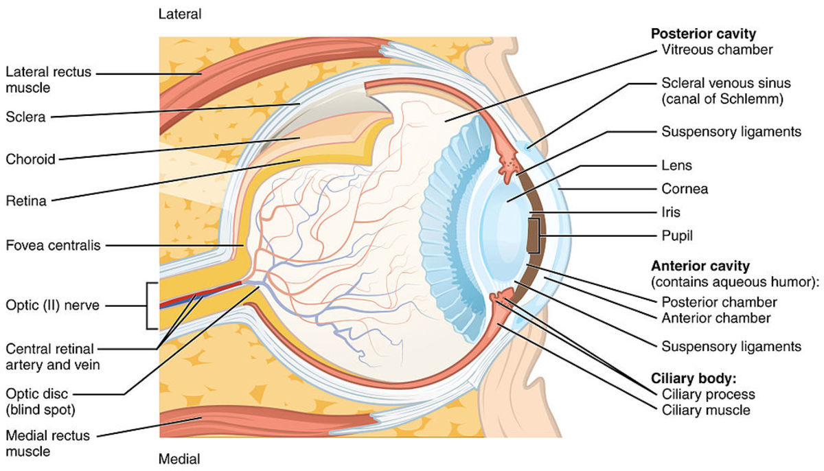
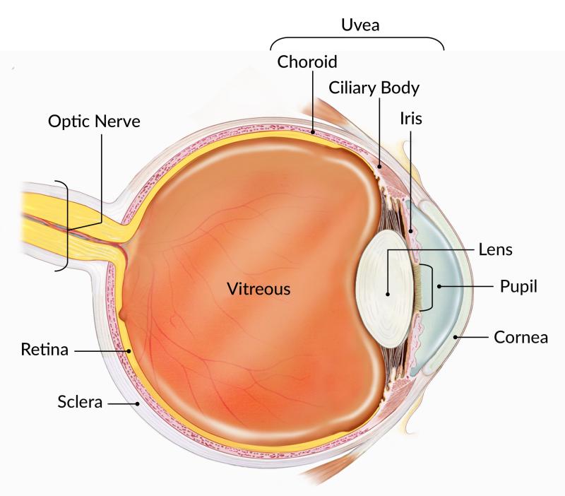
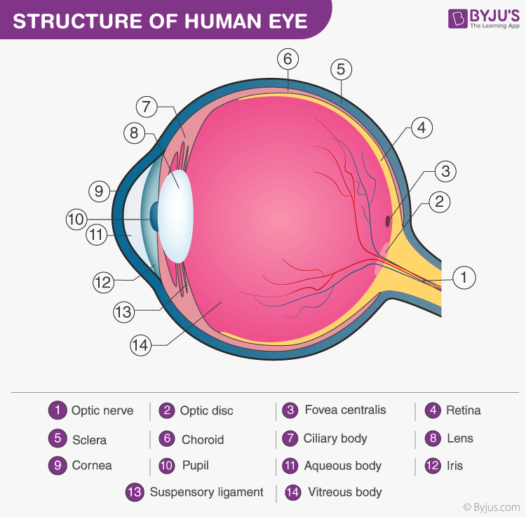
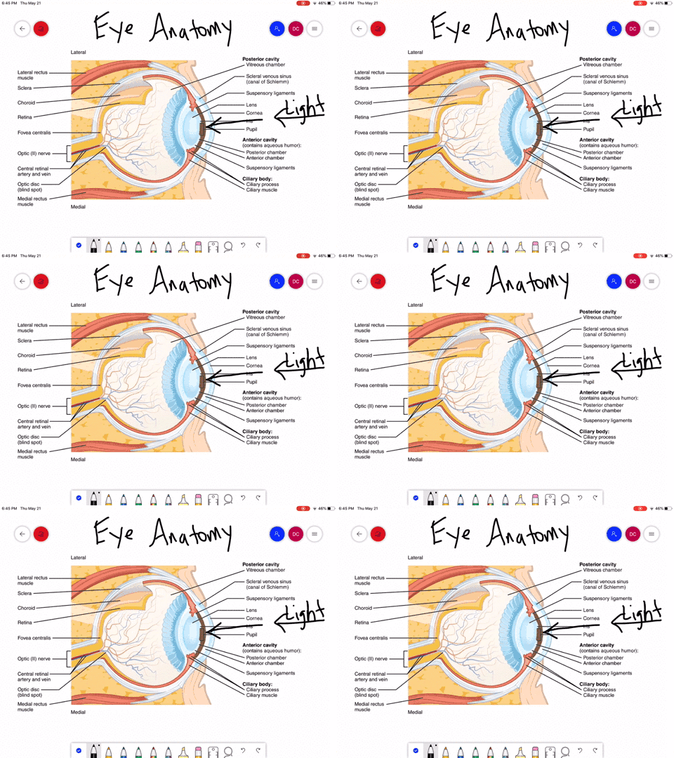
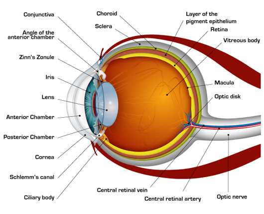
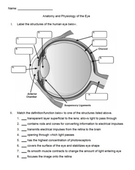
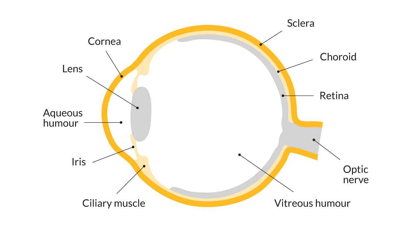
Eye Diagram With Labels and detailed description - BYJUS A brief description of the eye along with a well-labelled diagram is given below for reference. Well-Labelled Diagram of Eye. The anterior chamber of the eye is the space between the cornea and the iris and is filled with a lubricating fluid, aqueous humour. The vascular layer of the eye, known as the choroid contains the connective tissue.

Eye diagram with labels and functions
Anatomy of the eye: Quizzes and diagrams | Kenhub Take a look at the diagram of the eyeball above. Here you can see all of the main structures in this area. Spend some time reviewing the name and location of each one, then try to label the eye yourself - without peeking! - using the eye diagram (blank) below. Unlabeled diagram of the eye. Click below to download our free unlabeled diagram of ... Labelling the eye — Science Learning Hub Labelling the eye — Science Learning Hub Activity Labelling the eye The human eye contains structures that allow it to perceive light, movement and colour differences. In this activity, students use online or paper resources to identity and label the main parts of the human eye. Citizen science Teacher PLD Glossary Sign in Email Us Microscope, Microscope Parts, Labeled Diagram, and Functions A microscope is a laboratory instrument used to examine objects that are too small to be seen by the naked eye. It is derived from Ancient Greek words and composed of mikrós, "small" and skopeîn,"to look" or "see". It is one of the most revolutionized scientific instruments used to observe or examine minute structures not clearly ...
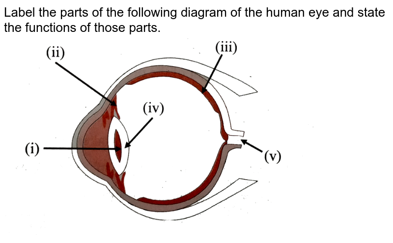
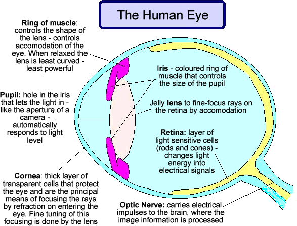
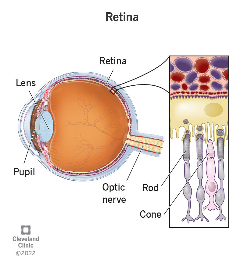
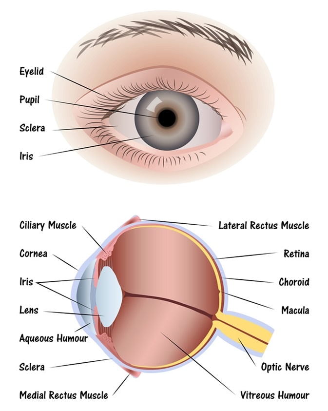
Eye diagram with labels and functions. Draw a labeled diagram of human eye. Write the functions of Cornea ... Cornea of the eye is the first sight where convergence of light rays takes place. Iris is that part of the eye which controls the amount of light entering the eye through the pupil. Pupil is a type of small hole through which light enters the eye. The eye lens is a convex lens just behind the pupilwhich converges the light rays towards the ratina. Microscope Types (with labeled diagrams) and Functions Simple microscope labeled diagram Simple microscope functions It is used in industrial applications like: Watchmakers to assemble watches Cloth industry to count the number of threads or fibers in a cloth Jewelers to examine the finer parts of jewelry Miniature artists to examine and build their work Also used to inspect finer details on products Labelled Diagram of Human Eye, Explanation and Function - VEDANTU The basic functions of Rods and Cones are conscious light perception, color differentiation and depth perception. The human eye is capable of distinguishing between about 10 million colors, and it can also detect a single photo. The human eye is a part of the sensory nervous system. Labeled Diagram of Human Eye Eye Diagram - an overview | ScienceDirect Topics (a) A perfect square eye diagram, (b) a closed eye diagram due to bandwidth, fiber impairments, noise, and timing jitter. The Q-factor ( q) is also an important system parameter widely used in long-distance optical transmission system design. It is defined as the electrical signal-to-noise ratio before the decision circuit at receiver.
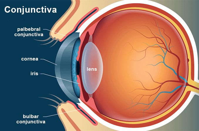
Eye Anatomy: A Closer Look At the Parts of the Eye - All About Vision Despite this, many people don't have a good understanding of the anatomy of the eye, how vision works, and health problems that can affect the eye. Read on for a basic description and explanation of the structure (anatomy) of your eyes and how they work (function) to help you see clearly and interact with your world. How the eye works Eye diagram basics: Reading and applying eye diagrams - EDN Eye diagrams provide instant visual data that engineers can use to check the signal integrity of a design and uncover problems early in the design process. Used in conjunction with other measurements such as bit-error rate, an eye diagram can help a designer predict performance and identify possible sources of problems. Also see: eye labeling Diagram | Quizlet conjunctiva delicate membrane lining the inside of the eyelids and covering the eyeball cornea fibrous transparent layer of clear tissue like a dome that covers the anterior portion of the eyeball (the iris and pupil). It is the first structure to refract (bend) light that enters the eye. sclera Tough white out covering of the eyeball choroid Rudyard.org | Eye anatomy diagram, Eye anatomy, Diagram of the eye Iridology. Simple eye diagram | Easy eye diagram | Labeled eye diagram We provide you Simple eye diagram and easy eye diagram from exam point of view. Also labeled eye diagram and anatomy of eye and human eye structure for better understanding. Human eye diagram and functions with diagram of human eye with labelling.
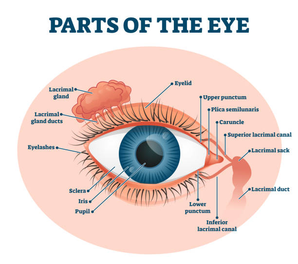
Human Eye Ball Anatomy & Physiology Diagram - eMedicineHealth The orbit is the bony eye socket of the skull. The orbit is formed by the cheekbone, the forehead, the temple, and the side of the nose. The eye is cushioned within the orbit by pads of fat. In addition to the eyeball itself, the orbit contains the muscles that move the eye, blood vessels, and nerves. The orbit also contains the lacrimal gland ... Eye Anatomy Diagram - EnchantedLearning.com Our eyes are organs that let us see. Eyes detect both brightness and color. Having two eyes separated on our face enables us to have depth perception (the ability to see the world in three dimensions - 3D). How we see: A whole series of events happens in order for us to see something. First, light must reflect off an object. Structure and Function of the Eyes - Eye Disorders - Merck Manuals ... The front section ( anterior segment) extends from the inside of the cornea to the front surface of the lens. It is filled with a fluid called the aqueous humor, which nourishes the internal structures. The anterior segment is divided into two chambers. The front (anterior) chamber extends from the cornea to the iris. Parts of a microscope with functions and labeled diagram - Microbe Notes Head - This is also known as the body. It carries the optical parts in the upper part of the microscope. Base - It acts as microscopes support. It also carries microscopic illuminators. Arms - This is the part connecting the base and to the head and the eyepiece tube to the base of the microscope.
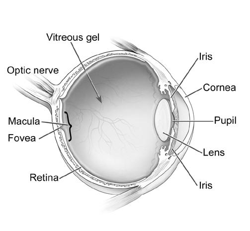
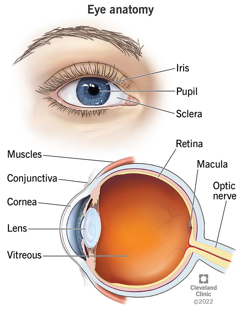
PDF Parts of the Eye - National Institutes of Health Iris: The iris is the colored part of the eye that regulates the amount of light entering the eye. Lens: The lens is a clear part of the eye behind the iris that helps to focus light, or an image, on the retina. Macula: The macula is the small, sensitive area of the retina that gives central vision. It is located in the center of the retina.
Cow's Eye Dissection - Eye diagram - Exploratorium A clear, flexible structure that makes an image on the eye's retina. The lens is flexible so that it can change shape, focusing on objects that are close up and objects that are far away. A muscle that controls how much light enters the eye. It is suspended between the cornea and the lens. A cow's iris is brown.
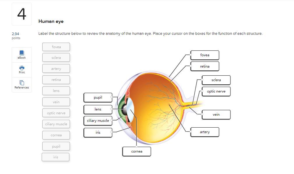
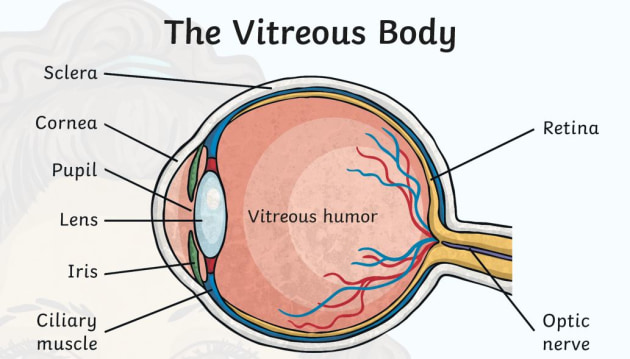
Labelling the eye — Science Learning Hub Labelling the eye Interactive Add to collection Use this interactive to label different parts of the human eye. Drag and drop the text labels onto the boxes next to the diagram. Selecting or hovering over a box will highlight each area in the diagram. Optic nerve Lens Schlera Pupil Vitrous humour Iris Cornea Retina Download Exercise Tweet
Structure and Function of the Human Eye - ThoughtCo To understand how the eye sees, it helps to know the eye structures and functions: Cornea: Light enters through the cornea, the transparent outer covering of the eye. The eyeball is rounded, so the cornea acts as a lens. It bends or refracts light. Aqueous Humor: The fluid beneath the cornea has a composition similar to that of blood plasma.
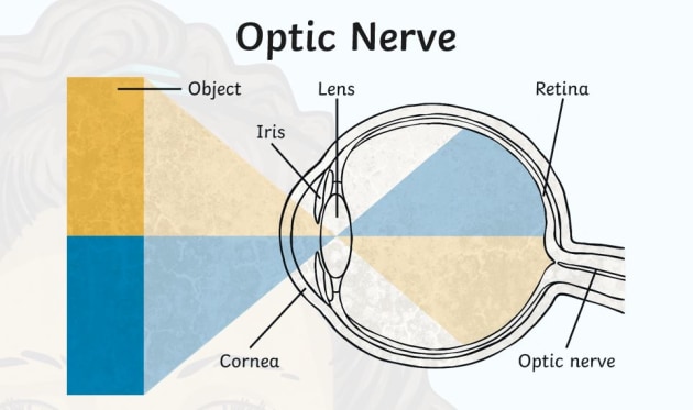
The Human Eye - Diagram, Parts, Working, Function and Work of The Lens The cornea, iris, pupil, and lens make up the front of the eye, which focuses the image onto the retina. The light-sensitive membrane that covers the back of the eye is known as the retina. This membrane is made up of millions of nerve cells that clump together behind the eye to form the optic nerve, a huge nerve. The Human Eye
What Does the Eye Look Like? - Diagram of the Eye | Harvard Eye Associates Fovea: A tiny pit located in the macula of the retina that provides the clearest vision of all. Iris: The colored part of the eye that controls the amount of light that enters the eye by changing the size of the pupil. Lens: A crystalline structure located just behind the iris - it focuses light onto the retina.
PDF Eye Anatomy Handout - National Institutes of Health Pupil: The pupil is the opening at the center of the iris. The iris adjusts the size of the pupil and controls the amount of light that can enter the eye. Retina: The retina is the light-sensitive tissue at the back of the eye. The retina converts light into electrical impulses that are sent to the brain through the optic nerve. Vitreous gel:
Parts of the Eye Labelled Diagram PowerPoint - CfE - Twinkl This fantastic PowerPoint of eye diagrams for kids is ideal for teaching CfE Second Level learners about the different parts of the eye and their functions. The PowerPoint takes pupils through each part of the eye with detailed labelled diagrams, and provides key information on how our eyes work and how we see.The PowerPoint begins by asking learners to consider how amazing our eyes are ...
Diagram of the Eye - Lions Eye Institute The eye - one of the most complex organisms in the human body. It is made up of many different parts working in unison together. In order for the eye to work at its best, all parts must work well collectively. To understand the eye and its functions, it's important to understand how the eye works, see below diagrams for both the external ...
Eye Anatomy: 16 Parts of the Eye & Their Functions - Vision Center The following are parts of the human eyes and their functions: 1. Conjunctiva. The conjunctiva is the membrane covering the sclera (white portion of your eye). The conjunctiva also covers the interior of your eyelids. Conjunctivitis, often known as pink eye, occurs when this thin membrane becomes inflamed or swollen.
Structure and Functions of Human Eye with labelled Diagram - BYJUS It keeps our eyes moist and clear and provides lubrication by secreting mucus and tears. Cornea: It is the transparent, anterior or front part of our eye, which covers the pupil and the iris. The main function is to refract the light along with the lens. Iris: It is the pigmented, coloured portion of the eye, visible externally. The main function of the iris is to control the diameter of the pupil according to the light source.
Microscope, Microscope Parts, Labeled Diagram, and Functions A microscope is a laboratory instrument used to examine objects that are too small to be seen by the naked eye. It is derived from Ancient Greek words and composed of mikrós, "small" and skopeîn,"to look" or "see". It is one of the most revolutionized scientific instruments used to observe or examine minute structures not clearly ...
Labelling the eye — Science Learning Hub Labelling the eye — Science Learning Hub Activity Labelling the eye The human eye contains structures that allow it to perceive light, movement and colour differences. In this activity, students use online or paper resources to identity and label the main parts of the human eye. Citizen science Teacher PLD Glossary Sign in Email Us
Anatomy of the eye: Quizzes and diagrams | Kenhub Take a look at the diagram of the eyeball above. Here you can see all of the main structures in this area. Spend some time reviewing the name and location of each one, then try to label the eye yourself - without peeking! - using the eye diagram (blank) below. Unlabeled diagram of the eye. Click below to download our free unlabeled diagram of ...



















Post a Comment for "45 eye diagram with labels and functions"